
- #advances-in-cardiovascular-imaging-and-diagnostics - main-overview
- #evolution-of-cardiac-imaging - history-and-progress
- #non-invasive-techniques - safer-approaches
- #ct-and-mri - detailed-heart-visualization
- #ultrasound-and-echocardiography - real-time-assessment
- #nuclear-cardiology - molecular-imaging
- #ai-and-machine-learning - future-of-diagnostics
- #case-studies - real-world-examples
- #patient-benefits - faster-and-safer-diagnosis
- #clinical-integration - challenges-and-opportunities
- #expert-guidance - heartcare-hub-support
Advances in Cardiovascular Imaging and Diagnostics: A Closer Look
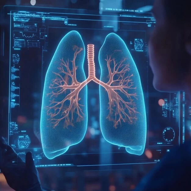
The phrase advances in cardiovascular imaging and diagnostics represents one of the most significant shifts in modern medicine. Decades ago, heart disease could only be diagnosed after major symptoms appeared. Today, cardiologists can visualize blood flow, detect structural changes, and even predict risk before critical events occur. This transformation is driven by technology, data, and clinical experience working hand in hand. For patients seeking guidance on the latest tools and services, HeartCare Hub provides access to resources, expert consultations, and tailored product recommendations.

1) The Evolution of Cardiac Imaging
From X-rays to high-definition scans
Cardiac imaging began with basic chest X-rays, offering little more than silhouettes of the heart. Over time, angiography, ultrasound, and nuclear scans pushed the field forward. Each innovation made heart disease more visible and diagnosable, allowing for earlier interventions.
Northside Hospital Cardiovascular Institute - Sandy Springs, Barfield
northside cardiovascular institute
6135 Barfield Rd Suite 100, Sandy Springs, GA 30328, USA

Historical note
In the 1970s, invasive catheter angiography was considered the “gold standard.” Today, patients can undergo non-invasive CT angiography in minutes, with lower risks and faster results.
2) Non-Invasive Techniques: A New Standard
Why less is more
Non-invasive cardiovascular imaging has become the preferred approach for both patients and clinicians. Techniques like cardiac CT, MRI, and echocardiography provide detailed insights without requiring invasive catheterization. This reduces complications, speeds recovery, and improves patient comfort.
3) CT and MRI in Heart Imaging
Precision and clarity
Cardiac CT scans allow physicians to detect plaque buildup in coronary arteries, often before symptoms appear. MRI, on the other hand, excels at identifying tissue damage after a heart attack or inflammation in myocarditis cases. Together, they provide a comprehensive map of heart health.
Case insight
One Cleveland patient avoided unnecessary surgery after an MRI revealed that her chest pain was linked not to coronary blockages but to inflammation, changing the treatment plan entirely.
4) Ultrasound and Echocardiography
Real-time assessment
Echocardiography remains a cornerstone in cardiology. It offers real-time visualization of heart valves, chamber sizes, and pumping strength. Doppler ultrasound further tracks blood flow, detecting regurgitation or stenosis with precision.
5) Nuclear Cardiology and Molecular Imaging
Looking beyond structure
Nuclear imaging, including PET and SPECT scans, reveals how well the heart muscle receives blood and oxygen. Molecular imaging goes a step further, detecting early changes in cellular activity before physical damage occurs—helping clinicians predict disease progression.
6) AI and Machine Learning in Diagnostics
The next frontier
Artificial intelligence is revolutionizing cardiovascular diagnostics. Algorithms now assist radiologists in identifying subtle abnormalities in scans, cutting interpretation times and reducing human error. AI-driven predictive models also help stratify patient risk more accurately than traditional methods.
Real-world example
A 2022 study showed that AI-enhanced echocardiograms detected early heart failure 20% faster than standard review alone, allowing for earlier medication adjustments.
7) Case Studies: Real-World Examples
From research to patients
In Boston, a middle-aged man underwent a CT angiogram after routine screening. The scan revealed significant coronary plaque despite no outward symptoms, leading to timely lifestyle interventions. In another case, AI-assisted MRI in London prevented misdiagnosis of myocarditis as anxiety, proving how critical accurate imaging can be.
8) Patient Benefits of Modern Imaging
Why patients win
Today’s cardiovascular imaging advances mean faster diagnosis, reduced invasiveness, personalized treatment, and improved survival rates. Patients not only gain more years of life but also better quality of life, as conditions are detected and treated earlier.
9) Clinical Integration: Challenges and Opportunities
Barriers to full adoption
Despite remarkable progress, challenges remain: equipment costs, training requirements, and access disparities between urban and rural healthcare systems. However, as technologies become more affordable and widespread, integration will only deepen.
10) Expert Guidance and Trusted Support
Where to find reliable help
Navigating the evolving world of cardiovascular imaging can be overwhelming. Partnering with experts ensures patients benefit from the latest techniques without unnecessary confusion. HeartCare Hub offers trusted information, professional consultations, and recommendations on services that align with each patient’s unique needs.


CardioVascular Group Lawrenceville
cardiovascular group
2200 Medical Center Blvd ste 400, Lawrenceville, GA 30046, USA